Dens evaginatus is a phenomenon that many people often call teeth growing sharp spikes. You can detect it with the naked eye. However, this condition should be handled appropriately to avoid future risks.
What’s dens evaginatus?
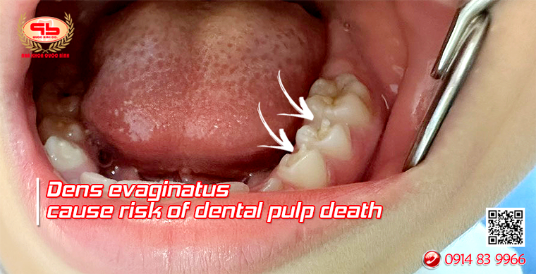
On the tooth, there is a separate protrusion in the form of a nub, a cusp. This extra nub has all the parts of a normal tooth such as enamel, dentin and pulp tissue inside.
Dens evaginatus can appear on the inside of the upper molars, or the chewing surface of the lower molars.
We can easily observe and detect this difference.
Cause
Dens evaginatus is caused by the inner enamel epidermis turning outward. However, there is also an opinion that this condition is genetic. Or due to local trauma affecting the tooth germ.

Potential risks
Because the dens evaginatus on chewing surface has a similar structure to a normal tooth but is protruding above the chewing surface. This leads to the pulp tissue protruding, making the tooth susceptible to wear and tear, exposing the pulp during chewing.
The chewing force at the dens evaginatus position will be stronger than at other positions. It is the first point of contact between the two jaws when moving, which can lead to complications and dental diseases over time. For example, pulp necrosis, periapical abscess, and painful swelling of the gums at the base of the molar.
In addition, in children 9-10 years old; Teeth with dens evagibatus hinder the bite, creating pressure on the tooth and root tip. Easily traumatized, causing the tooth to not close the apex, pulpitis, and root inflammation.
Treatment direction when encountering a dens evaginatus
A dens evaginatus can be treated by grinding and adjusting to completely remove it. And the first requirement is to take a dental X-ray. This grinding needs to be done properly and depends on many different factors. For example, the thickness of the dentin enamel layer on the pulp horn, the patient’s sensitivity. Otherwise, it can expose the pulp and cause pain.
@ If it does not affect the bite
In this case, no intervention is needed. Just need to prevent tooth decay in the groove between the secondary cusp and the tooth surface
@ If it affects the bite
The treatment will be to gradually reduce the height of the tooth cusp to regenerate the dentin on the pulp surface. Use Fluoride to remineralize the enamel surface, grinding repeatedly. Each time is 3 to 6 months apart to reduce the height of the secondary cusp and let the dentin gradually re-form
The treatment process is as follows:
-Use a fine-grained diamond drill at high speed with water spray to grind down the height of the secondary cusp.
-Then apply Fluoride to remineralize.
– Grinding with different appointments, each time the amount of grinding will depend on the thickness of the dentin layer above. The interval between two grinding adjustments is 3 months, monitor and re-evaluate after 12 months.
-Control within 1 year, grinding until the bite paper test shows that there is no longer any obstruction to the bite.
-In case grinding causes pulp exposure, perform direct pulp capping with biological filling material (MTA or Biodentin) and continue to monitor.
So if there are extra buds on the tooth, pay attention:
The extra buds are small and seemingly harmless, but can lead to unpredictable consequences.
When detecting extra buds on the tooth, absolutely do not grind or break them yourself.
When detecting such extra buds, go to the dentist to be examined and treated properly.
—  —
—
QUOC BINH DENTAL CLINIC (formerly VUNG TAU)
General Director – Dr. PHAN QUOC BINH
Add 1: 19 Pham Hong Thai, Tam Thang Ward, Ho Chi Minh City.
09148399 66/ (0254) 383 99 66
ADD 2: 28 Le Loi, Vung Tau Ward, Ho Chi Minh City.
(0254) 381 83 18 / 0942 231 212
Add 3: 649 Truong Cong Dinh, Tam Thang Ward, Ho Chi Minh City
Doctor in charge: Dr NGUYEN HUU CHIEN
0708 649 649
Working hours: 7:30-11:30, 14:00-20:30
https://nhakhoaquocbinh.com/en/price-list/
https://www.facebook.com/nhakhoathammyquocbinh